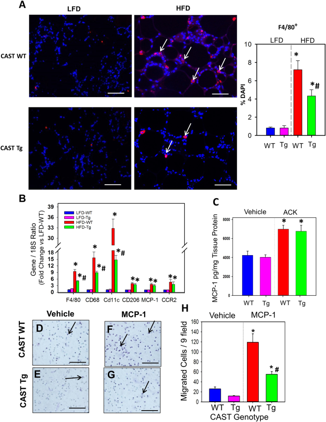
Figure 4.
CAST overexpression significantly reduced macrophage accumulation. (A) Representative immunofluorescent staining of F4/80 in EpiWAT cross-sections from 16 week LFD and HFD fed CAST WT and Tg mice. The nuclei were stained with DAPI (blue) and the F4/80-positive cells (red) are indicated by arrows. Using fluorescent microscopy, F4/80-positive cells were counted from 10 fields at the power of 100x magnification (n = 5). (B) mRNA abundance of F4/80, CD68, CD11c, CD206, MCP-1 and CCR2 genes in EpiWAT from LFD and HFD fed CAST WT and Tg mice were analyzed by qPCR (n = 5). (C) MCP-1 protein accumulation in adipose tissue explant culture media was measured by ELISA. (D-G) WT and CAST-Tg BMDMs were seeded on transwell filters and lower chambers were filled with media containing either vehicle or MCP-1 (100 µg/mL). Cells that migrated through the membrane stained with hematoxylin and were counted from 9 fields at the power of 200x magnification (H) (n = 4). Values are represented as mean ± SEM. *and # denotes P < 0.05 when comparing LFD vs HFD and WT vs Tg respectively (Two-way ANOVA with Holm-Sidak post hoc analysis).

