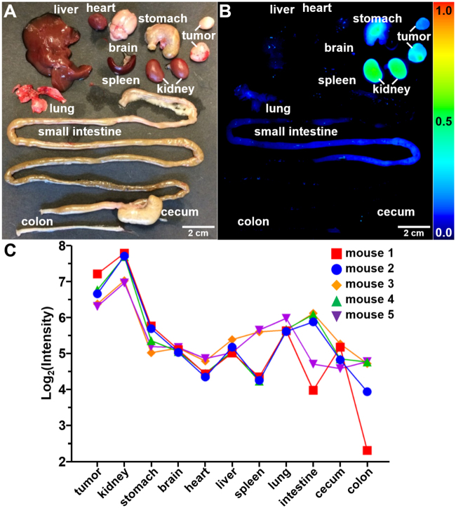
Figure 5.
Peptide biodistribution. (A) White light image shows individual organs from tumor-bearing mouse euthanized 1 hour after injection of KSP*-IR800, including human breast cancer xenografts (tumors), liver, heart, stomach, brain, lung, spleen, kidney, small intestine, cecum, and colon. (B) Fluorescence image shows high peptide uptake in tumors by comparison with other organs. Strong signal from kidneys support renal clearance. (C) Quantified fluorescence intensities are shown for tumor and other organs from n = 5 mice. We fit a two-way ANOVA model to log-transformed data with terms for 11 tissues and 5 mice and tested each tissue mean against the mean for tumors. Kidney had 1.7 times more intense signals on average (P = 0.03), while all other tissues had at least 2-fold lower signals than tumor on average (P = 0.008 for lung was the largest P-value).

