Figure 6.
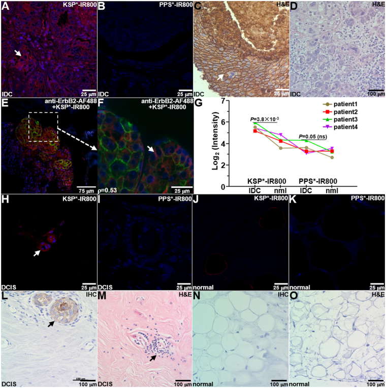
Specific peptide binding to human breast cancer ex vivo. (A) On immunofluorescence (IF), KSP*-IR800 shows strong staining to the surface (arrow) of invasive ductal carcinoma (IDC) cells from human specimens that express ErbB2, while minimal signal is seen with control (B) PPS*-IR800. (C) Immunohistochemistry (IHC) with anti-ErbB2 antibody confirms results. (D) Representative histology (H&E) for IDC. (E) Binding by KSP*-IR800 peptide (red) and anti-ErbB2-AF488 antibody (green) co-localizes on ErbB2 + IDC specimen with Pearson’s correlation coefficient of ρ = 0.53. (F) Magnified region from dashed box in (E) shows cell surface staining (arrow). (G) Quantitative comparison of KSP*-IR800 and PPS*-IR800 binding to human IDC (ErbB2+) with normal (ErbB2−) breast tissue. We fit an ANOVA model with terms for 4 conditions and 4 patients to log-transformed data and found a 2.44-fold greater signal for KSP*-IR800 in IDC than normal, but only a 1.30-fold increase for the same comparison with PPS*-IR800 peptide. The difference of differences was not significant, P = 0.09, which results from small number of human specimens. (H) On IF, we observed good staining of KSP*-IR800 to the surface (arrow) of non-invasive human ductal carcinoma in situ (DCIS) and (I) minimal signal with PPS*-IR800. On IF, we observed minimal staining to normal human breast tissue with either (J) KSP*-IR800 or (K) PPS*-IR800. (L) On IHC, we found strong reactivity to the surface (arrow) of DCIS cells. (M) Representative histology for DCIS. (N) Minimal reactivity was seen for normal human breast on IHC. (O) Representative histology for normal breast.

