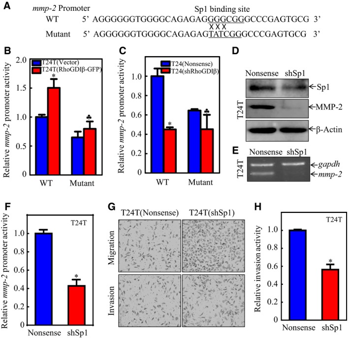
Figure 4.

Sp1 contributed to mmp‐2 transcription and further invasion of BC cells. (A) Illustration of the mutated Sp1 binding site in the mmp‐2 promoter region. (B,C) Wild‐type or mutant mmp‐2 promoter‐driven luciferase reporters was co‐transfected together with pRL‐TK into (B) T24T(Vector) and T24T(RhoGDIβ‐GFP) cells or (C) T24(Nonsense) and T24(shRhoGDIβ) cells. Twenty‐four hours post‐transfection, the transfectants were extracted to evaluate the luciferase activity. The results were presented as mmp‐2 promoter activity relative to the scramble vector transfectant transfected with wild‐type mmp‐2 promoter‐driven luciferase reporter, and each bar indicates mean ± SD from three independent experiments. *Significant difference in comparison to scramble vector transfetant. ♣Significant difference in comparison with wild‐type reporter transfectant (P < 0.05). (D, E) The cell extracts from T24T(Nonsense) and T24(shSp1) transfectants were subjected to western blot to determine (D) MMP‐2 protein expression or (E) mmp‐2 mRNA levels. (F) Wild‐type mmp‐2 promoter‐driven luciferase reporter was co‐transfected together with pRL‐TK into T24T(Nonsense) and T24T(shSp1) cells, respectively. Twenty‐four hours post‐transfection, the transfectants were extracted to evaluate the luciferase activity. The results were presented as mmp‐2 promoter activity relative to the vector control transfectants; each bar indicates mean ± SD from three independent experiments. *Significant difference (P < 0.05). (G,H) Invasion abilities of T24T(shSp1) and T24T(Nonsense) cells were evaluated using BD BioCoat™Matrigel™ Invasion Chamber (G). The relative invasion activity was plotted. The bars are mean ± SD from three independent experiments. *Significant difference between T24T(shSp1) and T24T(Nonsense) cells (P < 0.05).
