Figure 7.

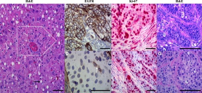
Histological and immunohistochemical evaluation of (A) liver and (B–G) tumor tissue samples. Hematoxylin and eosin (H&E) staining (A) showed single‐cell necrosis (white arrows) and necrosis of small groups of hepatocytes (white box); that is, moderate damage of liver tissues isolated from mice administered with the highest single dose of HisDianthin‐EGF (40 μg). (B) High EGFR expression level (arrow) was observed in untreated tumors, whereas (C) a weak expression of EGFR (arrow) was observed in the treatment group depicting curative effect of therapy as compared to untreated. High Ki67 proliferation index (D) was observed in all tumors except the one obtained after combination therapy [50%; (E)]. The right row of photographs (F, G) represents H&E‐stained samples corresponding to areas of the immunohistologically EGFR‐ and Ki‐67‐stained samples. Part (F) shows the tumor of an untreated mouse, whereas part (G) depicts the tumor of the sole mouse that retained a tumor after treatment with HisDianthin‐EGF + SO1861. The immunohistochemical samples are counterstained by hematoxylin. All scale bars represent a length of 100 μm.
