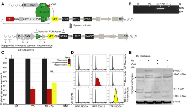
Figure 3.

In vitro activation of the oncogene cassette through CAG‐Flp transfection. (A) Schematics of Flp‐mediated activation of oncogene cassette. Arrows indicate primers for the transition PCR assay (1–2) and for the three ddPCR assays; pig genome (3–4), oncogene cassette (5–6), and recombination (7–8); (B) validation of recombination in Flp‐transfected TG fibroblasts using the transition PCR assay. The expected transition band of 972 bp is seen in Flp‐transfected TG fibroblasts, but not in untransfected, or WT fibroblasts; (C) ddPCR quantification of pig genomes, oncogene cassettes, and recombination events in WT, TG‐ and TG CAG‐Flp‐transfected fibroblasts. Plotted are mean ± SE, normalized to pig genome equivalents for three biological replicates; (D) flow cytometric assessment of BFP, RFP, and YFP fluorescence from WT, TG‐ and TG CAG‐Flp‐transfected pig fibroblasts (n > 10 000 cells in each analysis). All fibroblasts are BFP negative as expected. There is a complete shift from RFP to YFP in TG fibroblasts upon Flp transfection. Shown is a representative example of three independent experiments; (E) western blotting of untreated TG fibroblast, CAG‐Flp‐transfected TG fibroblasts with either DOX treatment or subsequent CAG‐Dre recombinase transfection. Anti‐2A linker peptide antibody was used to visualize multiple protein expression with β‐actin as loading control. The anti‐2A linker peptide antibody also recognizes various porcine teschovirus proteins, which results in a few unspecific bands in the immunoblot.
