Figure 3.
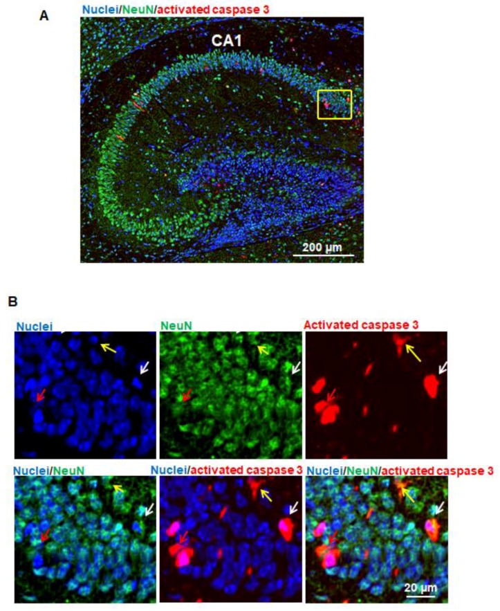
Propofol induces apoptosis in neurons in the hippocampus. (A) Fluorescence images of the hippocampus treated with propofol. In order to identify whether neurons in hippocampus undergo apoptosis following propofol exposure, the brain sagittal section were co-stained with activated caspase 3 and neuronal nuclear antigen (NeuN; neuron marker). Cell nuclei were stained with Hoechst 33342. Blue, green, and red represent cell nuclei, NeuN, and activated caspase 3 signals. Scale bar = 200 µm; (B) The magnified view of the yellow-boxed region shown in Figure 3A. These images include the individual channel and overlaid images and show that some activated caspase 3 and NeuN signals were co-localized in the same cells, suggesting that propofol induces apoptotic neuron death. Three representative apoptotic neurons are indicated by yellow, white, and red arrows. Scale bar = 20 µm.

