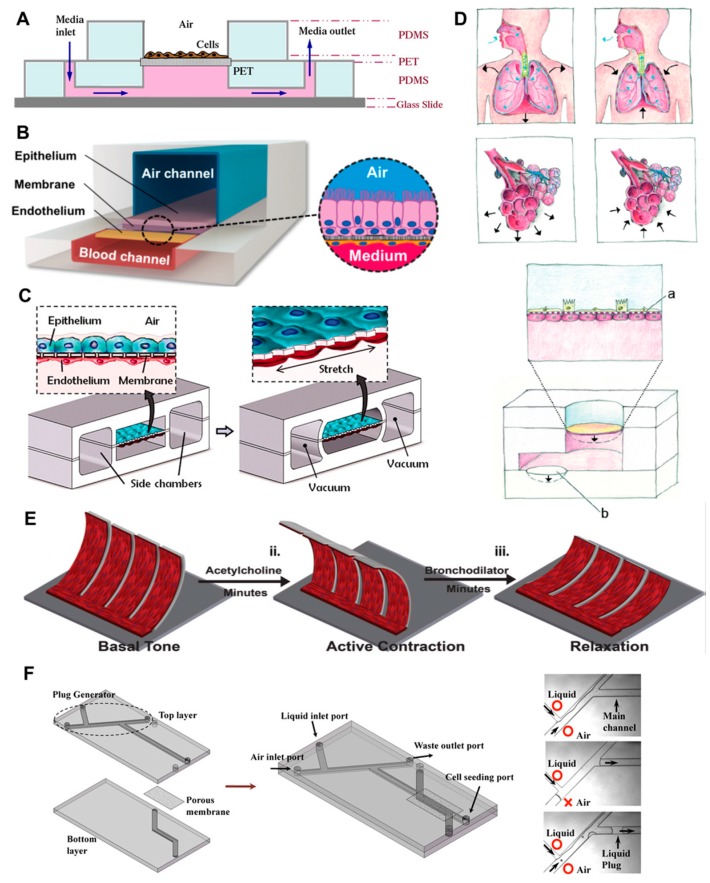
Figure 2.
Different lung-on-a-chip design principles. (A) The cross-sectional view though the microfluidic device shows an early attempt to recreate lung physiology by introducing an air–liquid interface to the cells. Reprinted from [150], with permission from Elsevier; (B) 3D cross-sectional view illustrates the use of an epithelial and endothelial cell line separated by a porous membrane that divides air and a media channel. Reprinted by permission from Macmillan Publishers Ltd: Nature Methods [151], copyright (2015); (C) Building on similar design considerations, the stretchability of the separating membrane is introduced by using vacuum chambers at both sides of the actual compartments for cell growth. From [154], reprinted with permission from AAA; (D) Since breathing introduces 3D multidirectional mechanical stress to the alveoli in human lung, a system was introduced that simulates this stretching more close than unidirectional stretching does. [156]—Published by The Royal Society of Chemistry under creative commons Attribution 3.0 Unported License; (E) Schematic depicting engineered bronchial smooth muscular thin films adopting (i) basal tone, (ii) preconstriction, and (iii) bronchodilator-induced relaxation. Reproduced from [158] with permission from The Royal Society of Chemistry; (F) Shown is a schematic of the microfluidic airway model and photomicrographs of the liquid plug generation. Reprinted from [160], © Springer Science+Business Media LLC 2011, with permission from Springer. PET: Polyethylene

