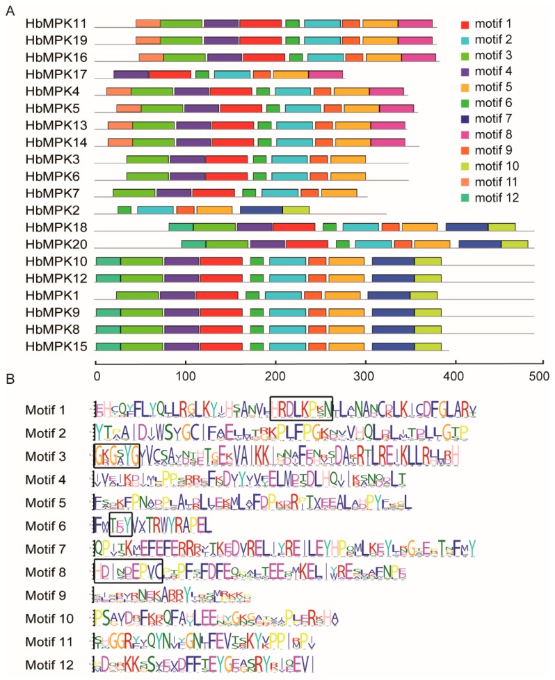
Figure 4.
Motif analysis of the MPK family members in Hevea. (A) Motif distributions for 20 HbMPKs. Rectangles with different colour represent 12 recognised conserved motifs; (B) Detailed information (amino acid sequence) of the 12 motifs. The heights of each letter represent the frequency of amino acids at that position, and the black boxes indicate the key sites of the four conserved motifs.

