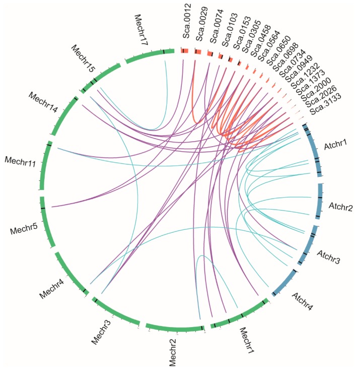
Figure 7.
Segmental duplication of HbMPK genes and syntenic analysis of Hevea, M. esculenta, and A. thaliana MPKs. Chromosomes and scaffolds are shown in different colour in circular form. The positions of the MPK genes are marked by black lines on the circle. Duplicated MPK pairs in Hevea are connected by red lines. Syntenic relationships between Hevea and the other two species are connected by purple lines. Blue lines indicate MPK pairs between or inside M. esculenta and A. thaliana.

