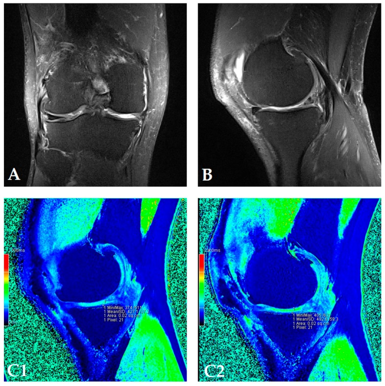
Figure 2.
(Patient dG07) Coronal (A) and sagittal (B) MRI show complete loss of articular cartilage of the medial femoral and tibial condyles (ICRS grade IV chondromalacia), thinning and shallow fissures of the articular cartilage in the lateral femoral and tibial condyles (ICRS grade IV chondromalacia), edge osteophytes and joint effusion. The MRI with the dGEMRIC index values at four-time points (T0: baseline; T3: three months after autologous and microfragmented adipose tissue injection; T6: six months after autologous and microfragmented adipose tissue injection; T12: 12 months after autologous and microfragmented adipose tissue injection) (C1–C4). The scheme of the dGEMRIC index with different joint facets throughout the study period at T0, T3, T6 and T12 combined with VAS scale ratings in T0, T3, T6, T12 (D).


