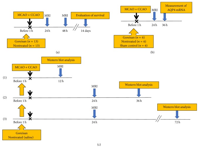
Figure 1.
Experimental procedure and design. (a) Rats were divided into Goreisan and nontreated groups. Rats in the Goreisan group received Goreisan (2.0 g/kg) dissolved in saline (total volume 400 μl) administered orally 1 h before surgery. Rats in the nontreated group received saline. MRI [T2-weighted (T2w) and diffusion-weighted (DW)] was performed at 24 and 48 h, and the survival rate was evaluated 14 days after surgery. The infarcted area at the maximum incised surface was observed using MRI (DW and T2w). We calculated the comparative amounts of the infarcted areas using the formula of Swanson et al. [22]. (b) An MRI was performed at 24 h, and the brain was removed at 36 h to evaluate AQP4 expression using immunoblotting and quantitative reverse-transcription PCR. (c-(1)) We examined AQP4 protein levels using western blot analysis at 12 h after MCAO + CCAO for the acute phase. (c-(2)) MRI was performed at 24 h and the brain was dissected at 36 h for the subacute phase. (c-(3)) MRI was performed at 24 h and the brain was dissected at 72 h for the chronic phase.

