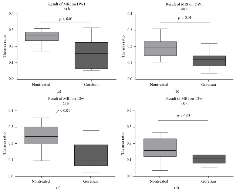
Figure 3.
Lesion area measured using MRI. Ratios of the ischemic area to the total slice area in MRI for (a, b) DWI and (c, d) T2w are expressed as the median, range, and 25–75 percentiles. The MRI findings indicated that the lesion areas were significantly smaller in the Goreisan group than in the nontreated group at 24 and 48 h.

