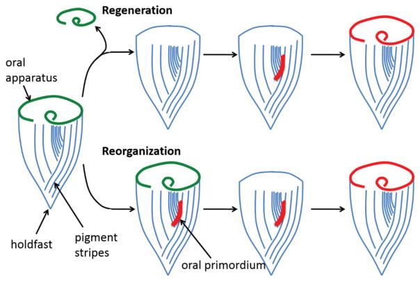
Figure 2.
The regeneration and reorganization of the oral apparatus (green) of Stentor coeruleus. Blue lines indicate surface pigment stripes, and the red region indicates the oral primordium. At the opposite end from the oral apparatus, Stentor possesses a posterior holdfast, which the cell uses to attach itself to a solid substrate. When an oral apparatus regenerates after removal, it first assembles as an oral primordium, which then migrates to the anterior end and becomes a new oral apparatus.

