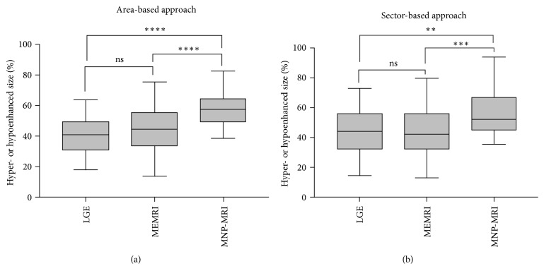
Figure 4.
Comparison of contrasted sizes of MI mice by different MRI methods. The sizes were evaluated by area-based (a) and sector-based approaches (b). Enhancement size of MNP-MRI was significantly larger than that of LGE and MEMRI for both approaches. Difference in the enhancement size between LGE and MEMRI was not significant for both approaches. ∗∗P < 0.01, ∗∗∗P < 0.001, ∗∗∗∗P < 0.0001, ns = nonsignificant. (area-based: P = 0.34 for LGE versus MEMRI, P < 0.0001 for LGE versus MNP-MRI and P < 0.0001 for MEMRI versus MNP-MRI; sector-based: P = 0.99 for LGE versus MEMRI, P = 0.0015 for LGE versus MNP-MRI and P = 0.0001 for MEMRI versus MNP-MRI).

