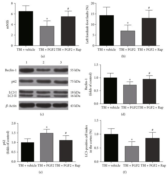
Figure 7.
Rapamycin reversed the beneficial effects of FGF2 on neurological function and changed the expression of autophagy-related proteins in the ipsilateral cortex 48 h post-TBI. (a, b) The quantification of mNSS and the percentage of left forelimb foot faults at 48 h after TBI induction. The bars represent the mean ± SD. n = 24. ∗P < 0.05 versus TBI + vehicle and #P < 0.05 versus TBI + FGF2. (c) Representative Western blots showing levels of Beclin-1, p62, and LC3 in the ipsilateral cortex 48 h post-TBI. Lane 1, TBI + vehicle group; lane 2, TBI + FGF2 group; lane 3 TBI + FGF2 + Rap group. (d, e) The relative band densities of Beclin-1 and p62. The densities of the protein bands were analyzed and normalized to β-actin. The data are expressed as a percentage of the sham group. The bars represent the mean ± SD. n = 6. ∗P < 0.05 versus TBI + vehicle, #P < 0.05 versus TBI + FGF2. (f) The band density ratio of LC3 II to LC3 I. The bars represent the mean ± SD. n = 6. ∗P < 0.05 versus TBI + vehicle and #P < 0.05 versus TBI + FGF2.

