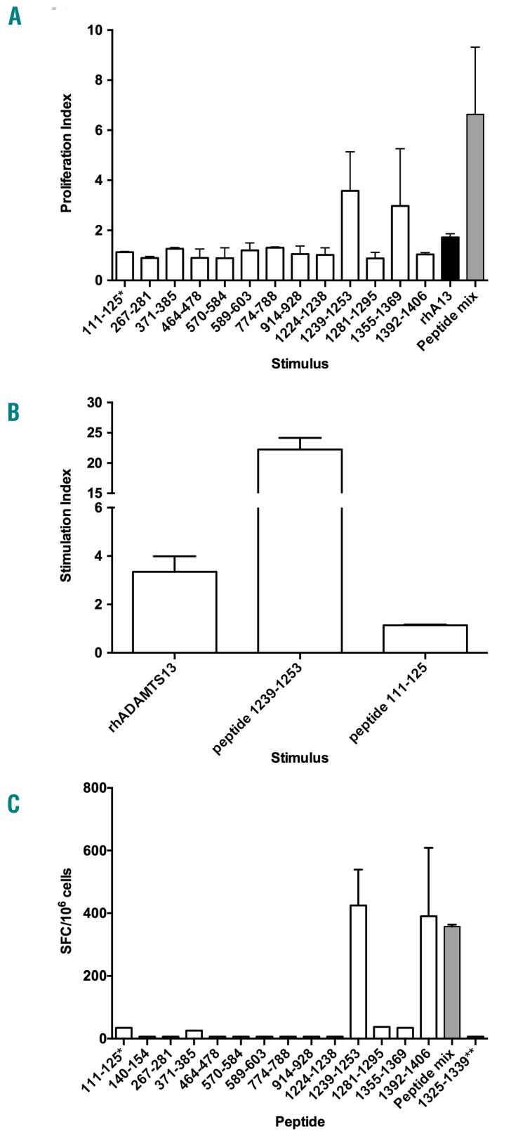
Figure 2.
The ADAMTS131239–1253 peptide is an immunodominant T-cell epitope both for Sure-L1 mice as well as for a patient with TTP. (A) Stimulation by ADAMTS13-derived peptides of spleen and lymph node cells from Sure-L1 mice immunized with rhADAMTS13. Cells (3×10⁵/well) were incubated with each individual peptide (10 μg/mL), with the pool of peptides (Peptide mix) or with rhADAMTS13. Proliferation was assessed by incorporation of tritiated thymidine. Proliferation indices are defined as the ratio of cpm of stimulated cells versus cpm of control cells, and are expressed as means±SD for two Sure-L1 mice. (B) The ADAMTS131239–1253 peptide is processed and presented to T cells by human antigen-presenting cells. A representative ADAMTS13-specific CD4+ T-cell hybridoma (clone 1F5γ) was incubated with immature dendritic cells from a healthy HLA-DR1+ blood donor and with the ADAMTS131239–1253 peptide, with rhADAMTS13 or with the ADAMTS13111–125 peptide. Stimulation indices represent the ratio of secretion of IL-2 by T cells stimulated in the presence of antigen versus the secretion of IL-2 by T cells incubated with Mo-DC alone. Means±SD are from two independent experiments. Similar results were obtained with other ADAMTS13-CD4+ T-cell hybridoma clones (not shown). The peptide incubated alone or with DR15+ artificial antigen-presenting cells failed to activate the T cells (not shown). (C) ADAMTS13 epitope specificity of CD4+ T-cell lines from a patient with acquired TTP. CD4+ T-cell lines were generated after stimulation with pooled ADAMTS13-derived peptides as defined in Table 1. T cells (3×10⁵/well) were then incubated with AAPCDR1 (3×10⁴ cells/well) alone or pulsed with individual peptide (10 μg/mL), or with the peptide pool (Peptide mix). The number of cells producing interferon-γ was then assessed by ELISPOT and is expressed as the mean number of spot-forming cells (SFC) per million cells calculated in the case of peptide-stimulated T cells minus the SFC obtained in the case of unstimulated cells. Means±SD are from two independent experiments. *The 111–125 ADAMTS13-derived peptide that did not bind to HLA-DRB1*01:01 was used as a negative control.

