Figure 2.
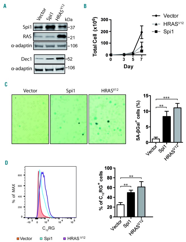
Overexpression of Spi1 and HRASV12 in Lin−Kit+Sca1+ (LSK) cells leads to senescence. (A) Western blot analysis of Spi1, HRASV12 and the senescence marker Dec1 in hematopoietic cells subjected to the retroviral-mediated expression of Spi1, HRASV12 or an empty vector. Protein extracts of GFP-positive sorted cells were analyzed 7 days post-infection as described in Online Supplementary Figure S2. α-adaptin served as the loading control. (B) Number of total living cells retrovirally transduced with Spi1 and HRASV12 or an empty vector (control), at the indicated periods of time. The means ± SEM of at least 3 independent experiments are shown. (C) Representative SA-βgal staining and mean percentages of SA-βgal positive cells (histograms) in samples of hematopoietic cells subjected to the retroviral-mediated expression of Spi1, HRASV12 or an empty vector as a control. SA-βgal assays for sorted GFP-positive cells were performed 7 days post-infection as described in Online Supplementary Figure S2. The counting of GFP-positive SA-βgal cells was performed in 9 randomly selected fields with a total of more than 2000 cells from each group. Magnification of images, 200X. The means ± SD of at least 3 independent experiments are shown. **P<0.005; ***P<0.0005 from two-tailed Student’s t-test. (D) Flow cytometric detection of SA-βgal activity using C12RG as a fluorogenic substrate in cells retrovirally transduced with Spi1, HRASV12 or an empty vector. The histograms represents the % of C12RG positive cells among GFP-positive cells. The means ± SD of at least 3 independent experiments are shown. **P<0.005 from two-tailed Student’s t-test.

