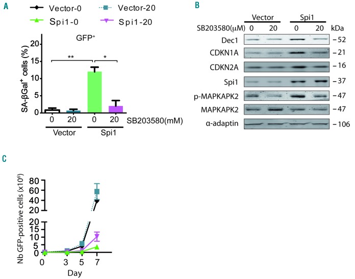
Figure 5.
Spi1-induced senescence requires P38MAPK14 signaling in hematopoietic cells. (A) Mean percentage of SA-βgal positive cells (histograms) subjected to the retroviral-mediated expression of Spi1 or an empty vector, and maintained in cultures with or without 20 μM of SB203580 in samples of GFP-positive sorted hematopoietic cells 7 days post-infection. The means ± SD of at least 3 independent experiments are shown. *P<0.05; **P<0.005 from two-tailed Student’s t-tests. (B) Hematopoietic cells transduced with empty vectors (vector) or Spi1 expression vectors and maintained in cultures with or without 20 μM of SB203580 were sorted for GFP-positive cells 7 days post-infection and subjected to Western blot analyses of CDKN1A, CDKN2A, p-MAPKAPK2, MAPKAPK2, Dec1 and Spi1. α-adaptin served as the loading control. (C) Number of GFP-positive cells retrovirally transduced with empty vectors (vector) or Spi1 expression vectors and maintained in cultures with or without 20 μM of SB203580 from day 1 to day 6 at the indicated periods of time. The means ± SD of at least 3 independent experiments are shown.

