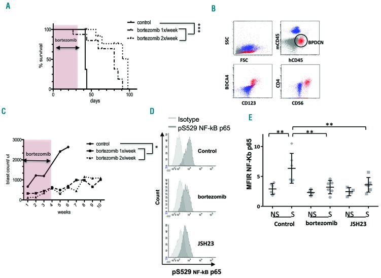
Figure 4.
Bortezomib treatment is efficient at controlling tumor growth in a xenograft model using primary blastic plasmacytoid dendritic cell neoplasm cells. NSG mice were irradiated (2 Gy) and then inoculated intravenously with 1×10⁶ to 2×10⁶ primary BPDCN cells from patient #127 on day 0. Treatment was started on day 100 (J1) after the graft with bortezomib (0.25 mg/kg/mouse intraperitoneally) given one or twice weekly for 4 weeks (n=7 and n=4 mice, respectively). Mice injected with phosphate-buffered saline (PBS) over the 4 weeks were used as the control (n=3). (A) Overall survival of BPDCN inoculated-mice treated with bortezomib (dotted line) or with PBS (solid line) is shown. (B) One example of the immunostaining of peripheral blood performed at day 89 after engraftment. Murine cells (blue) and primary BPDCN (red) cells are distinguishable based on specific human or murine CD45 antibody expression. Human BPDCN cells express CD123, BDCA4, and CD4. (C) Mean of BPDCN cell counts in the blood of mice following treatment with bortezomib (dotted line) or PBS (solid line). (*P<0.05 and ***P<0.001). Intracellular expression of pRelA (pS529 NF-κB p65) was evaluated in PDX cells (BPDCN patient #127) obtained in mouse blood at day 1 and day 15 after in vivo treatment with bortezomib (0.25 mg/kg/mouse intraperitoneally) for 6 h (n=3 mice). JSH23 was used as a positive control (40 mg/kg, n=3 mice) and PBS (control, n=3 mice) as a negative control. PDX cells were stimulated ex vivo with TLR7 for 45 min (R848, 1 μg/mL) before staining. (D) Representative examples of intracellular expression of pRelA and isotype control staining after ex vivo TLR7 stimulation in these different conditions. (E) This histogram represents the mean fluorescence intensity ratio (MFIR) ± SEM of intracellular NF-κBp-65 in PDX cells obtained after treatment with bortezomib on day 1 and day 15, *P<0.05, **P<0.01. NS: unstimulated; S: stimulated with R848. The MFIR was obtained by dividing the mean fluorescence intensity (MFI) obtained with the anti-NF-κBp-65 antibody by the MFI of the respective isotype control antibody.

