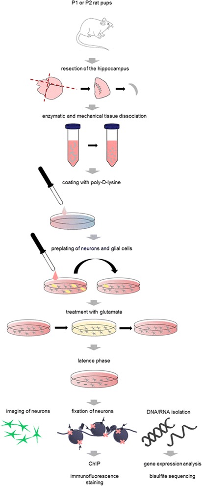
Fig. 1.

Experimental study design. Brains of P0 to P2 rat pups were removed, hippocampi resected and then enzymatically and mechanically dissociated. Culture dishes were coated with poly-D-lysine. Cell suspension was pre-plated for 1 h, to separate glial (yellow) and neuronal cell populations (grey) by sedimentation. Neuronal cell population was cultured on poly-D-lysine coated dishes. After 12 DIV neurons were treated with 10 μM glutamate for 10 min. Cells were further cultured for up to 4 weeks after glutamate injury and subsequently imaged, fixed with formalin or lysed for downstream applications
