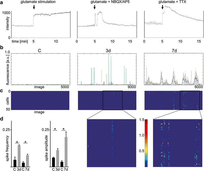
Fig. 3.

Development of spontaneous recurrent epileptiform discharges following glutamate injury in cultured rat hippocampal neurons. Calcium imaging. a Development of neuronal activity measured by calcium uptake and release of a single selected neuron before, during and after stimulation with glutamate and glutamate supplemented with either NBQX/AP5 or TTX. b Display of the development of synchronized neuronal activity determined by calcium imaging of 10 representative neurons 3 and 7 days after glutamate injury compared to sham control. c Heatmap showing exemplarily the intensity and frequency of calcium signals of 50 simultaneously recorded neurons 3 or 7 days after stimulation with glutamate. Insets highlight synchronization and bursting activity. d Mean of spike frequency and spike amplitude of recorded neurons 3 and 7 after glutamate injury is significantly increased compared to corresponding controls. All error bars represent standard deviation. Asterisks indicate significance (p < 0.05). AP5 - D-amino-5-phosphonovaleric acid; C – control; d – days; NBQX - 2,3-dihydroxy-6-nitro-7-sulfamoyl-benzo-quinoxaline-2,3-dione; TTX – tetrodotoxin
