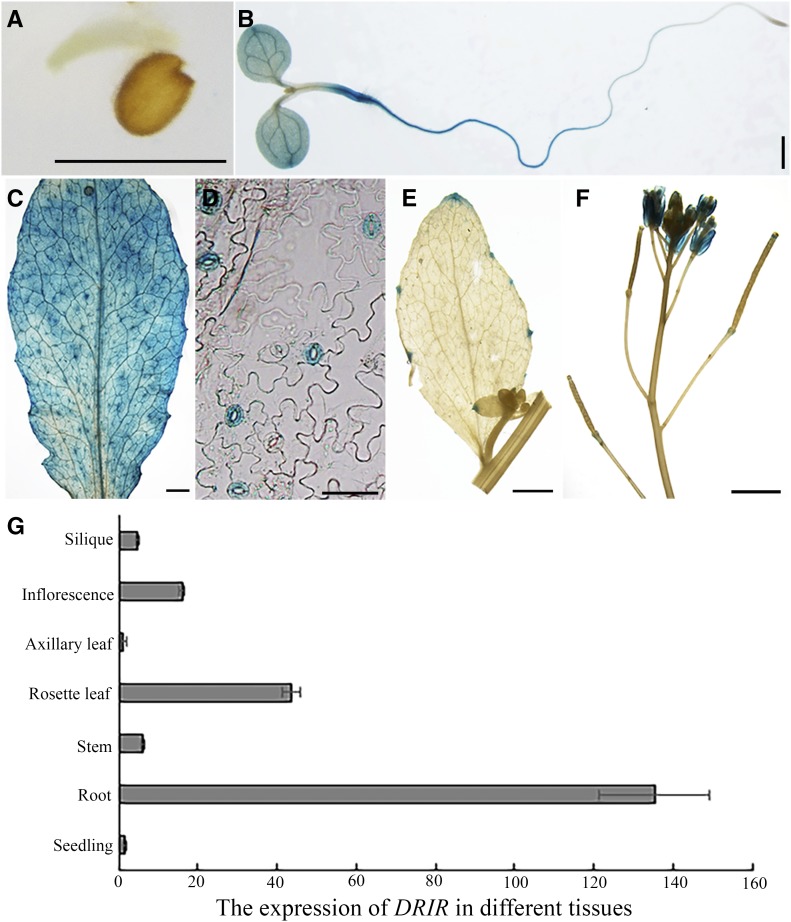
Figure 1.
Expression pattern of DRIR in seedlings. Shown are DRIR promoter-GUS activities in germinating embryo (A), 6-d-old seedling (B), rosette leaf (C), guard cells in rosette leaf epidermis (D), axillary leaf (E), and inflorescence (F). Bars = 1 mm (A, B, C, E and F) and 50 µm (D). G shows transcript abundance of DRIR in different tissues as determined by quantitative RT-PCR. Values shown are means ± sd from three biological replicates.

