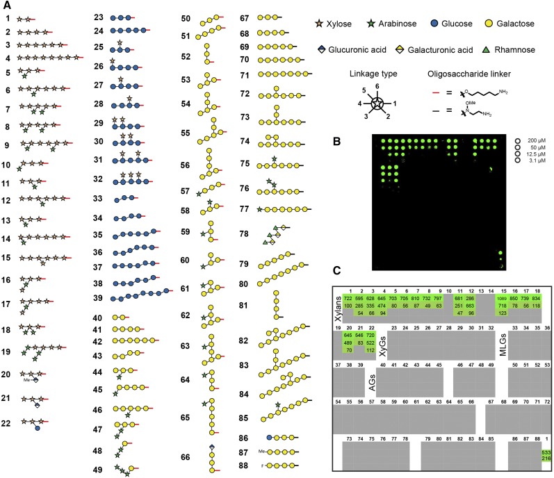
Figure 1.
A glycan microarray equipped with synthetic cell wall oligosaccharides. A, The printed oligosaccharides comprise fragments of four major polysaccharide classes: xylans (compounds 1–22), glucans (23–39), galactans (40–77 and 79–88), and rhamnogalacturonan (RG)-I (78). Red and black bars at the reducing end of the oligosaccharides indicate the different linkers of the respective compounds produced either by automated glycan assembly (1–66) or conventional solution-phase chemistry (67–88). The legend for linkage types denotes at which position the next monosaccharide is attached. B, Fluorescence signal for the binding of LM10 to xylan oligosaccharides. Each compound was printed at four different concentrations as indicated on the right. The printing pattern of the glycan microarray is depicted in C. C, Quantification of the fluorescence signal for LM10. The values denote fold change over background. Only signals of more than 4-fold above background and above 4% of the maximal value are shown. Note that the 200 and 50 µm concentrations of oligosaccharide 1 were reprinted on the array as the last spots (bottom right corner) to confirm constant printing efficiency.

