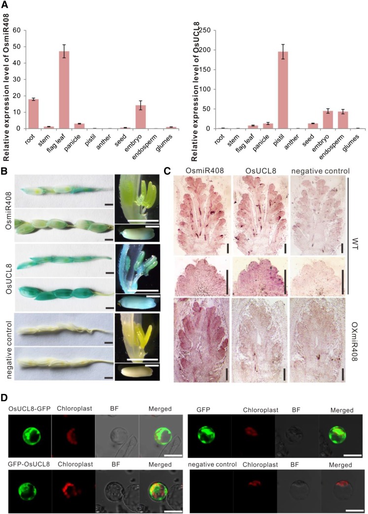
Figure 2.
Expression patterns of OsmiR408 and OsUCL8, and subcellular localization of the OsUCL8 protein. A, Relative expression patterns of OsmiR408 and OsUCL8 in different tissues. Values are expressed as means ± sd. B, Spatial expression pattern analysis of OsmiR408 and OsUCL8 in young panicles and grains by GUS staining. Bars = 2 mm. C, In situ hybridization of OsmiR408 and OsUCL8 during panicle development in wild-type (WT) plants and in OXmiR408 plants. Bars = 100 µm. D, Subcellular localization of OsUCL8. The empty vector with or without ATG before the GFP gene was used as a positive or negative control, respectively. BF, Bright field. Bars = 10 µm.

