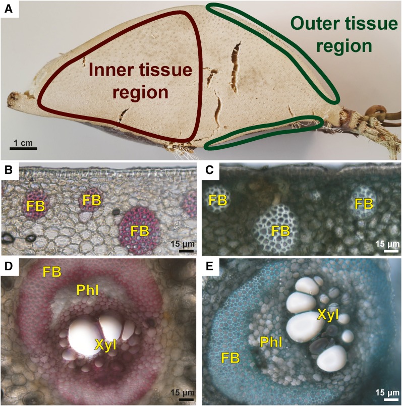
Figure 1.
Inner and outer tissue regions of the palm leaf base were sectioned and stained to show the presence of lignified walls. A, A thick, transverse segment of the leaf base used in this study showing the locations of the inner tissue and outer tissue regions. B, Image of a transverse section of the outer tissue region stained with phloroglucinol-HCl showing the red-stained, lignified walls of fibers present in fiber bundles (FB). C, Transverse section of the outer tissue region stained with Toluidine Blue O showing the thick, lignified, blue-stained walls of fibers present in fiber bundles. D, A vascular bundle in the inner tissue region stained with phloroglucinol-HCl. The bundle is surrounded by fibers, and there is a prominent fiber bundle cap over the phloem (Phl); the xylem tissue (Xyl) includes tracheary elements with lignified (red-stained) walls. The walls of the fibers surrounding the vascular bundle, including the cap, show only weak staining with phloroglucinol-HCl. The phloem cell walls were not stained and, therefore, are nonlignified. E, A transverse section of a vascular bundle similar to that in D showing blue staining of the fiber walls and blue-green staining of the tracheary element cell walls with Toluidine Blue O. Bars = 1 cm (A) and 15 μm (B–E).

