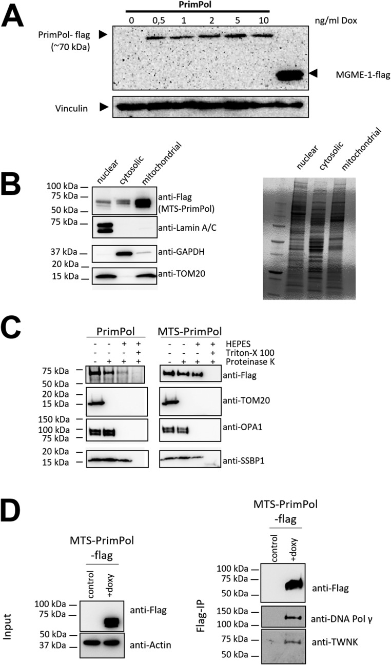
Fig. S7.
Mitochondrial localization of PrimPol and MTS-PrimPol. (A) Confirmation of Dox-dependent PrimPol-flag expression in Flp-InTM T-RExTM 293 cells. The cells were treated for 48 h with the indicated concentration of Dox in the growth medium. Recombinant PrimPol was detected from the total cell protein preparations with an anti-flag antibody, with MGME1-flag used as a positive control and vinculin as a loading control. (B) Mitochondrially targeted PrimPol is highly enriched in the mitochondrial fraction from induced MTS-PrimPol Flp-InTM T-RExTM 293 cells. (C) Mitochondrial fractions from PrimPol WT and MTS-PrimPol–overexpressing cells. MTS-PrimPol is exclusively mitochondrially localized as demonstrated by Proteinase K treatment of intact mitochondria, capable of digesting outer membrane proteins such as TOM20. When detergent (Triton X-100) is added, Proteinase K will also digest the mitochondrial proteins, including single-strand binding protein SSBP1. Note that the TFAM-targeting peptide is cleaved upon mitochondrial import and therefore does not influence the protein size. (D) Immunoprecipitation of mitochondrial-targeted PrimPol pulls down central mitochondrial replisome components, Pol γ and TWNK helicase. TWNK was also found to be a PrimPol interaction partner in the BioPlex study (33). Noninduced cells were used as the control input.

