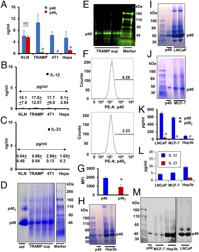
Fig. 1.
Levels of IL-12 family of cytokines in different cancer cells. Levels of p40 and p402 (A), IL-12 (B), and IL-23 (C) were measured in supernatants of mouse tumor cells (KLN, TRAMP-C2, 4T1, and Hepa) by ELISA. Results are mean ± SD of three different experiments (aP < 0.001 vs. p40 measured in respective tumor cells). (D) TRAMP-C2 supernatants were passed through a 10-kDa cut column followed by native PAGE and Coomassie blue staining. The p40 band was detected by comparing with pure p40 protein (far left column). (E) Native PAGE immunoblot analyses of p40 in the supernatants of TRAMP-C2 from three separate experiments. (F) Intracellular FACS assay of p40 and p402 in cultured TRAMP-C2 cells after live/dead cell exclusion. (G) Mean fluorescence intensity (MFI) of intracellular p40 and p402 was calculated (*P < 0.01 vs. p40; NS, not significant). Native PAGE followed by Coomassie staining was performed to detect p40 in supernatants of human cancer cells [Hep3B (H); LnCAP (I); MCF-7 (J)]. Levels of p40 and p402 (K) and IL-12 and IL-23 (L) were measured in supernatants of Hep3B, LnCAP, and MCF-7 cells by sandwich ELISA. Results are mean ± SD of three different experiments (aP < 0.001 vs. p40 measured in respective tumor cells). Native PAGE immunoblot analyses of p40 and other IL-12 members of cytokines (M) in supernatants of Hep3B, LnCAP, and MCF-7 cells.

