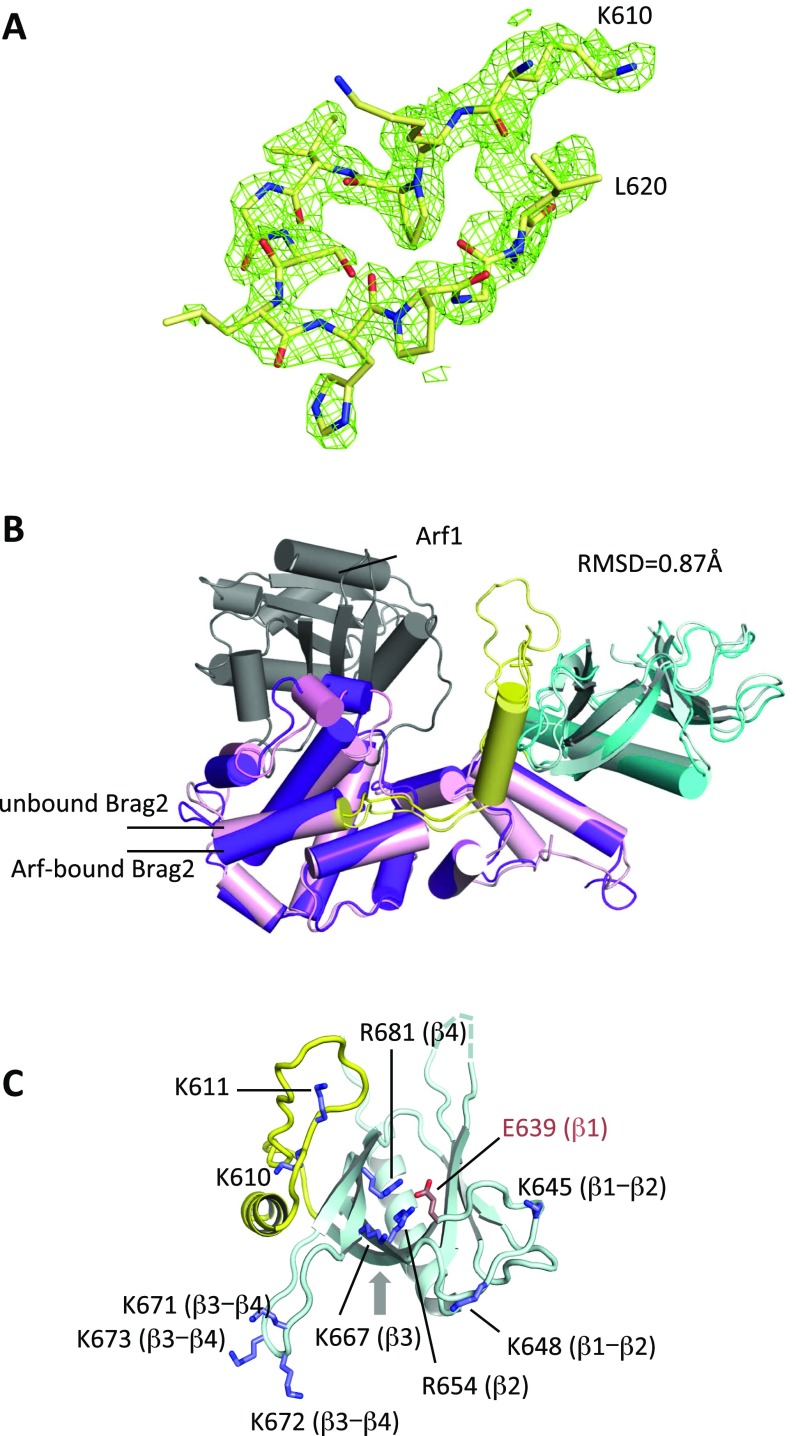
Fig. S1.
Crystallographic analysis of unbound Brag2Sec7-PH. (A) Omit map of the K610-L620 loop in the linker region (space group P212121). (B) Superposition of unbound Brag2 (present work, space group P212121, light colors) and Arf1-bound Brag2 (dark colors). The rmsd between the two structures is 0.87 Å. The color-coding of the Sec7, linker, and PH domains is as in Fig. 1A. Arf is shown in gray. The orientation is similar to that in Fig. 1A. (C) The membrane-facing surface of the linker and PH domains, showing exposed positively charged residues. The β-strand or the loops between β-strand to which each residue belongs is indicated. The alternative PIP2-binding site on the outer face of the β1-β2 strands is indicated by an arrow. The view is rotated by 90° with respect to Fig. 1A.

