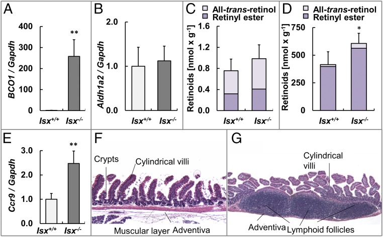
Fig. 1.
ISX-deficient mice display enlarged lymphoid follicles in the small intestine. (A and B) Quantitative RT-PCR analysis of jejunal Bco1 and Aldh1a2 mRNA levels normalized to Gapdh. (C and D) Levels of nonpolar retinoids (all-trans-retinol and retinyl ester) in jejunal (C) and hepatic (D) lipid extracts. (E) Quantitative RT-PCR analysis of jejunal Ccr9 mRNA levels normalized to Gapdh. (F and G) Representative H&E-stained cross sections through the small intestinal wall of a Isx+/+ (F) and a Isx−/− mouse (G). Values indicate mean ± SD of results from at least five animals per genotype. Threshold of significance was set at *P < 0.05 and **P < 0.01. (Magnification: 20×.)

