Fig. S5.
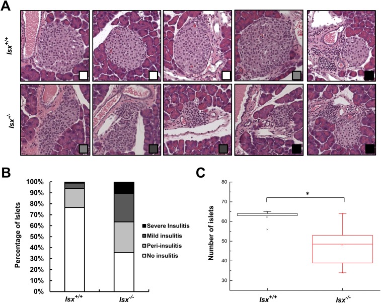
Pancreas morphology of Isx−/− and Isx+/+ mice. (A) H&E-stained cross sections of representative islets of Isx−/− and Isx+/+ mice. The degree of insulitis is indicated by the squares (bottom right corner) and refers to the legend displayed in B. (Magnification: 20×.) (B) Quantification of the histological appearance of islets of Isx−/− and Isx+/+ mice. Insulitis-scoring criteria were used as follows, and representative histology is shown in A: No insulitis (white); periinsulitis with infiltration of lymphocytes restricted to the periphery of the islet (light gray); mild insulitis with less than 50% of the islet area infiltrated by lymphocytes (dark gray); and severe insulitis with more than 50% of the islet area infiltrated with lymphocytes (black). (C) Total numbers of β-islets in pancreatic cross sections of Isx−/− and Isx+/+ mice. Values indicate mean ± SD of results from six animals per genotype. Statistical significance compared with the control group was assessed by ANOVA followed by Scheffé tests using software origin 9. Threshold of significance was set at *P < 0.05.

