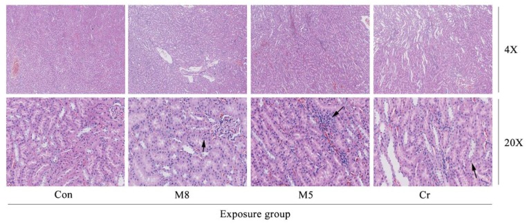
Figure 6.
Photomicrographs of kidney histopathology. The images (magnification 4×, 20×) represent the histopathological sections of the kidney. M8 group and Cr group (20×), large areas of renal tubular epithelial cells were cloudy and swollen, significant degeneration, vacuolar degeneration of the membrane (dark arrow). M5 group (20×) a large number of inflammatory cells infiltration (dark arrow). Abbreviations: Con, control group; M8: the mixture of eight common metals (Pb + Cd + Hg + Cu + Zn + Mn + Cr + Ni); M5: the mixture of five non-essential metals (Pb + Hg + Cd + Ni + Cr); Cr, chromium.

