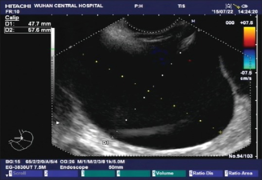
Figure 3.

Endoscopic ultrasound showed multiple anechoic round masses in the outer wall of the fundus and anterior wall of the stomach, with no internal blood flow signals. Partitions were visible between the masses, the sizes were approximately 75 mm × 47 mm, and the boundaries were consistent with liver imaging; hence, cysts were suspected
