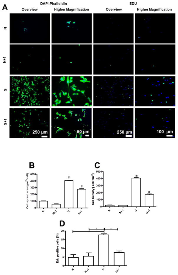
Figure 9.
(A) DAPI/phalloidin staining and representative images of EDU assay for HUVECs adhered on native films (N) and cross-linked films (G) with an additional bilayer on the top of the film and in the presence or absence of adsorbed or immobilized proteins. Quantification of (B) Cell number per field, (C) Cell spread area, and (D) Cell circularity. (E) Percentage of proliferating cells measured by the EDU assay. Statistical analysis was performed, and data was considered statistically different for p values < 0.05 (*). (#) denotes significant differences when compared to all PEMs formulations.

