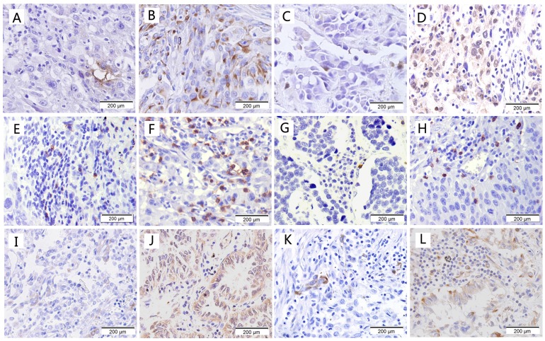
Figure 1.
Immunohistological staining of cancer stem cell markers and immune markers. (a) Low expression of CD133; (b) High expression of CD133; (c) Low expression of OCT-4; (d) High expression of OCT-4; (e) Low expression of CD8; (f) High expression of CD8; (g) Low expression of CD56; (h) High expression of CD56; (i) Low expression of HLA class I; (j) High expression of HLA class I; (k) Low expression of PD-L1; (l) High expression of PD-L1.

