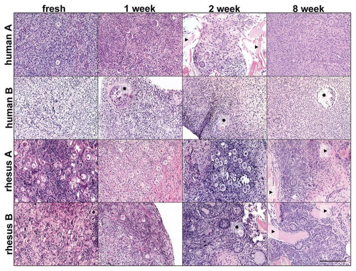
Figure 7.
Histological analysis of rhesus macaque and human ovarian cortical strip tissue cultured on OTP. Hematoxylin and eosin (H&E) stained sections of ovarian cortical tissue from two human and two rhesus macaque ovaries revealed multiple follicles within fresh tissue pieces and pieces cultured for one, two, and eight weeks. ★ = examples of multilayered growing follicles. ▶ = examples of OTP visible within tissue slice. Scale bar, 200 μm.

