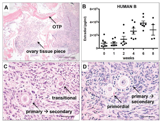
Figure 8.
Histological and secreted hormone analysis of human ovarian cortical strip tissue cultured on OTP. A) H&E stained sections of human ovarian cortical tissue cultured for two weeks on ovary tissue paper. Cells from the ovarian cortical tissue appear within the OTP pieces. B) The cortical strip pieces from human B were cultured on OTP for up to eight weeks. Estradiol was produced and secreted into the media over the course of this culture. Bars represent mean +/− SEM, n = 4 or 7 samples. C,D) H&E stained sections of human participant ovarian cortical tissue cultured for eight weeks. Ovarian follicles from multiple stages of differentiation are visible. Scale bar, (A,C,D) 50 μm.

