
Figure 1.
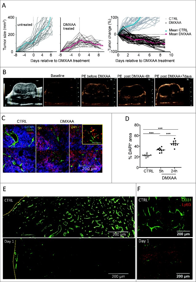
Anti-tumoral DMXAA effects begin with vascular damages and early cell death in transplanted PyMT tumors. A. Follow-up of transplanted PyMT tumor controls (left) and after one i.p. injection of DMXAA (23 mg/kg) (center). The right panel gives the relative tumor size changes after day 0, the day of treatment: [(size at day X)-(size at day 0)]/ (size at day 0). n = 20 tumors for each condition. The average curves are shown in color. B. Assessment of tumor perfusion using contrast ultrasound: from left to right: 1) acquisition in the largest diameter showing tumor anatomy; 2) baseline image in non-linear imaging mode before contrast enhancement of non-treated tumor; 3) Peak Enhancement (PE) image obtained after bolus injection of Ultrasound Contrast Agent (UCA) in the non-treated tumor; 4) UCA-PE image 6 hours after DMXAA injection; 5) UCA-PE image 7 d after DMXAA injection. Contrast enhancement observed in images after i.v. UCA injection illustrate that DMXAA treatment is associated with a strong alteration of vessel functionality as revealed by the absence of micro-bubbles circulation 6 h after DMXAA. Slight recovery of perfusion is observable at the tumor periphery after 7 d. C. DAPI staining shows an early DMXAA-induced cell death (DAPI+ cells) in both tumor and endothelial cells. D. Quantification of relative DAPI+ areas at different time points after DMXAA injection. Each point is a 20x image field, from n = 2 independent experiments. *** = p < 0.001 (t-test). E. As judged from CD31+ staining, 24 h after its i.p. injection, DMXAA has induced major vascular damages in the center of the tumor. The edge of the tumors is indicated with yellow lines. F. Ly6G+ neutrophils are much more abundant one day after DMXAA than in control conditions.