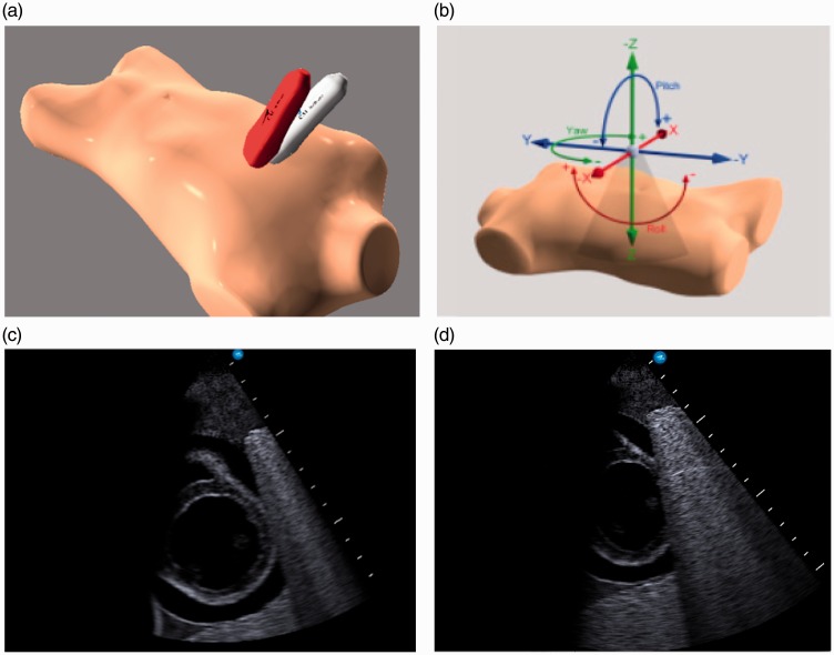
Figure 1.
Imaging on echocardiography simulator: (a) Reference probe position (red) vs. trainee image acquisition (grey); (b) Reference vs. trainee probe position difference measured in term of total angle difference (pitch + roll + yaw) and distance difference (x + y + z); (c) Example of reference image; (d) Example of trainee image

