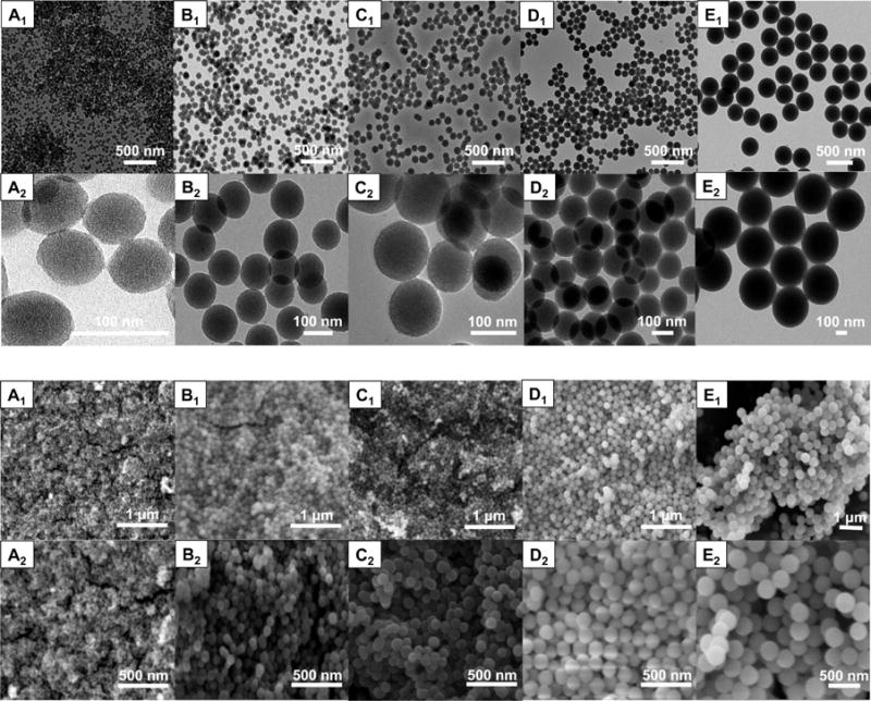
Figure 1.

TEM (top) and SEM (bottom) images of the nanoparticles at two magnifications. Nanoparticles from left to right: (A1, A2) 60 Mesoporous D; (B1, B2) 110 Mesoporous D; (C1, C2) 110 Mesoporous T; (D1, D2) 120 Nonporous D; and (E1, E2) 330 Nonporous D. Both TEM and SEM images indicate that highly uniform nanoparticles with low polydispersity were synthesized. On the basis of the images, nonporous nanoparticles had denser structure, with a smoother surface in larger nanoparticles.
