
Figure 7.
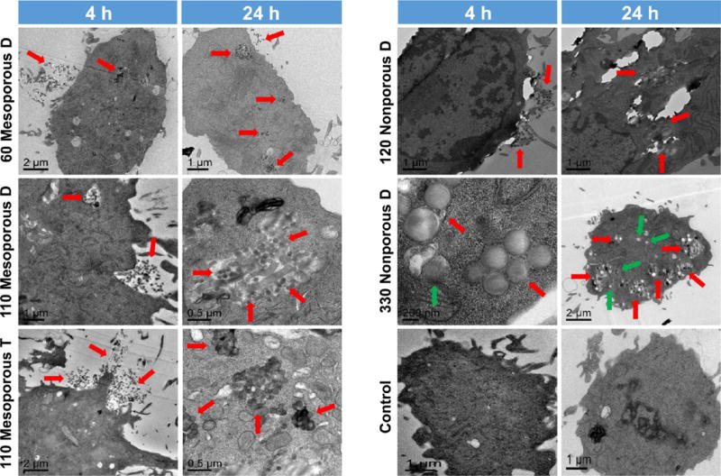
Cell uptake observed by TEM after 4 and 24 h of incubation of 80 μg mL−1 nanoparticles in RAW 264.7 macrophages. Nanoparticles were taken up in a time-dependent pattern and trapped in endosomal vacuoles. A few larger nanoparticles (especially 330 nm shown as green arrows) were isolated as individual nanoparticles.