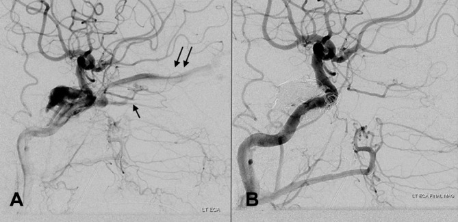
Figure 1.

(A) Lateral angiogram showing a carotid cavernous fistula (type D). Retrograde flow into the superior (double arrow) and inferior ophthalmic veins (single arrow). (B) Lateral angiogram after coil and Onyx embolisation confirming complete occlusion of the fistula.
