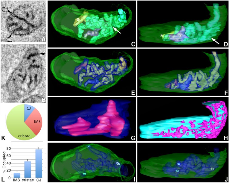
Fig. 5.
ET volumes of Mic19-miniSOG label similar domains in human primary astrocytes, including networks in the IMS. (A) A 1-nm slice through the center of a tomographic volume from an astrocyte showing Mic19-positive domains at CJs, IMS and also substantially in the intracristal space. CJs often showed a light open region (arrowheads) indicating low Mic19 occupancy in this small localized region. Results are representative of n=9 cells and 60 mitochondria. (B) Another example of a 1-nm slice through the center of a tomographic volume from a different astrocyte that has a darkly labeled Mic19-positive CJ (arrowhead). (C) The segmented and surface-rendered volume of the mitochondrion in B (OMM green, three cristae in various colors). There was one highly networked crista (arrow), and two much smaller and non-networked cristae (*). (D) As in C, but with a side orientation showing that the networked crista was far larger than the only two other cristae present. (E) A substantial portion of the cristae (translucent blue), typical of the 60 mitochondrial volumes examined, was occupied by the Mic19-positive domains (green). (F) Side orientation showing the Mic19-positive domains extending throughout the cristae and some of the cristae segments were filled with label. (G) One of the two smaller cristae showing a typical Mic19-positive domain inside that occupied about half the crista volume. The shapes of these domains were contiguous rather than speckled throughout. (H) IMS (magenta) Mic19-positive network against the IBM background (cyan). (I) Only three CJs (cyan) were present in this volume and are shown in relation to the OMM (translucent green) and cristae (translucent blue). (J) Side view showing the CJ openings. (K) Pie chart showing the distribution of Mic19-positive domains between CJ, IMS and cristae. (L) Histogram showing the percentage of the IMS, cristae or CJ volumes occupied by Mic19-positive domains (mean±s.e.m.). Results are from three biological replicates and 10 total technical replicates (K,L).

