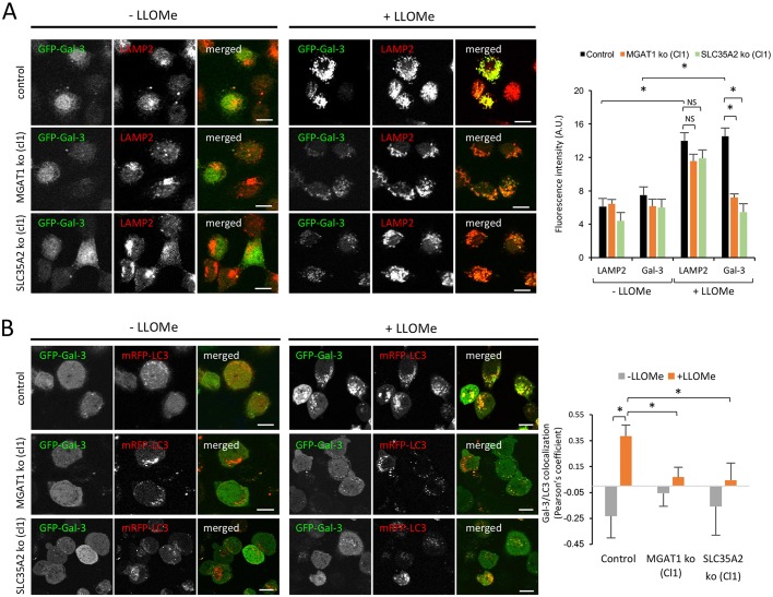
Fig. 6.
Recruitment of GFP-Gal3 to damaged lysosomes is reduced in MGAT1- and SLC35A2-deficient cells. (A) Wild-type (control), MGAT1-deficient (clone 1) or SLC35A2-deficient (clone 1) sHeLa transiently expressing GFP-Gal-3 for 24 h were treated with 1 mM LLOMe for 3 h. Cells were fixed with PFA, permeabilized with Triton X100 and subjected to immunocytochemistry using an anti-LAMP2 antibody, then processed for confocal microscopy. The intensity of LAMP2 and Gal-3 signals measured using ImageJ in a minimum of 20 cells per condition is shown on the right. (B) Wild-type (control), MGAT1-deficient (cl1) or SLC35A2-deficient (cl1) sHeLa transiently expressing GFP-Gal-3 and mRFP-LC3 for 24 h were treated with 1 mM LLOMe for 3 h. Cells were fixed with methanol and processed for confocal microscopy. Colocalization (Pearson's coefficient) between Gal-3 and LC3 is shown on the right. Data are mean±s.e.m. from individual cells (n>20); *P<0.05 (two-sample Student's t-test). Scale bars: 10 μm.

