Fig. 1.
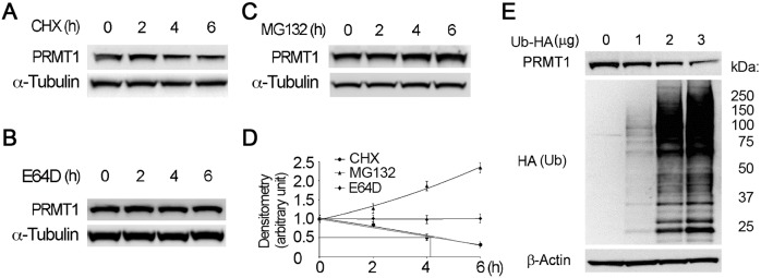
PRMT1 undergoes proteasomal degradation. (A–D) Cells were treated with (A) CHX (40 µg ml–1), (B) MG132 (20 µM) or (C) E64D (20 µM) for the indicated times, and cell lysates were subjected to immunoblotting analysis with antibodies against PRMT1 and α-tubulin. The relative PRMT1 protein levels from densitometry analysis of the immunoblots were plotted and the half-life of PRMT1 was calculated as previously described (Li et al., 2017) (D). (E) Cells were transfected with indicated amounts of HA-tagged ubiquitin plasmid, and the cell lysates were immunoblotted with anti-PRMT1, HA and β-actin antibodies. The results are representative of n=3 experiments.

