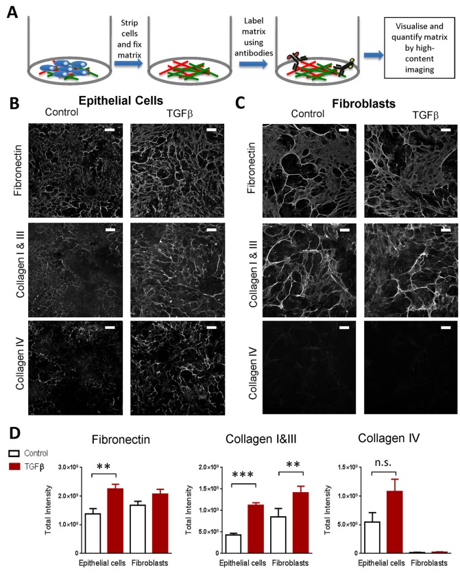
Fig. 2.
Stimulated proximal tubular epithelial cells produce a mature, deposited extracellular matrix comparable to that generated by fibroblasts. (A) Diagram showing the method for immunofluorescence analysis of deposited ECM. (B) Epithelial cells cultured for 6 days with or without stimulation with 10 ng/ml TGFβ1. Cells were then stripped, extracellular matrix fixed, and cells stained using antibodies against collagen I, III, IV and fibronectin. Each panel shows an image of one representative field from four independent experiments. Scale bars: 100 µm. (C) Fibroblasts cultured with or without stimulation for 6 days and stained as in B. Scale bars: 100 µm. (D) The graphs show the results of the quantification of fibrillary extracellular matrix from images as in B and C. Results are the mean from four independent experiments with 6 replicates per experiment, t-test performed on means from independent experiments, **P<0.01, ***P<0.001; error bars indicate mean±s.e.m.

