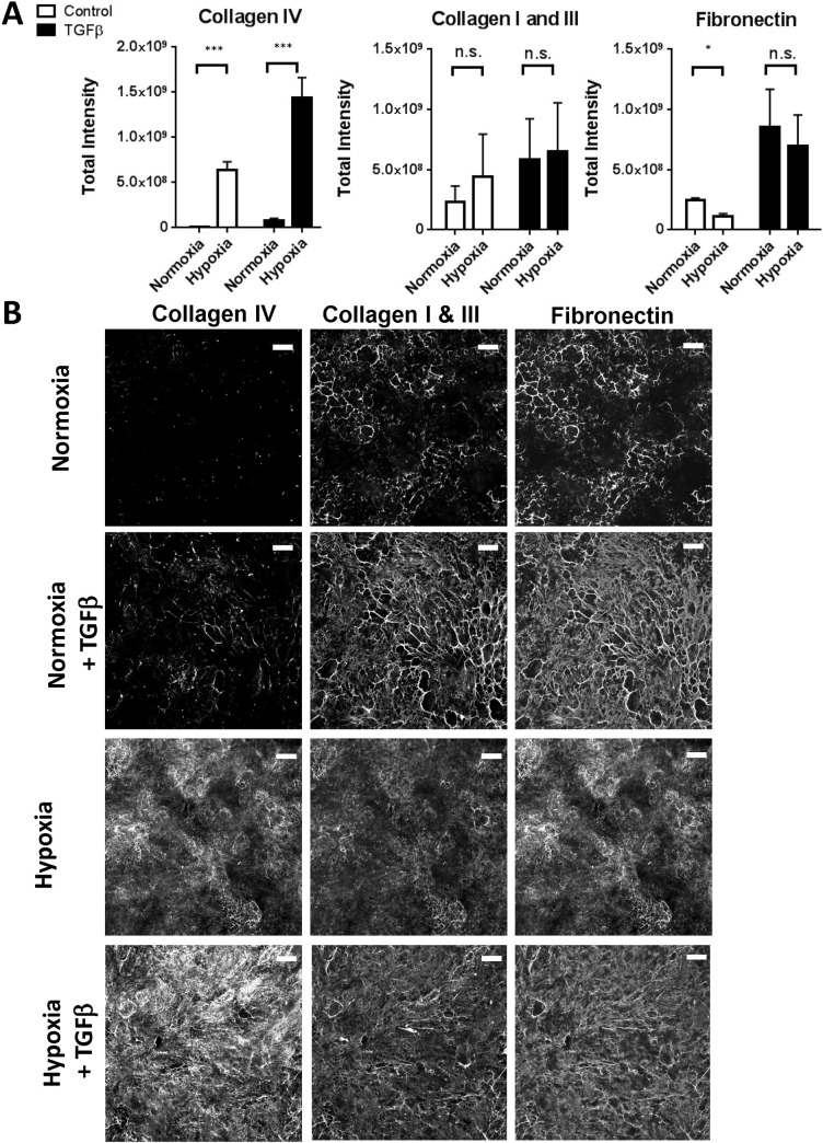
Fig. 5.
Response of RPTECs to hypoxia. (A) Epithelial cells grown for six days in either normoxic or hypoxic (2.5% O2) conditions with or without stimulation with 10 ng/mL TGF-β1 were assessed for extracellular matrix production using the immunofluorescence assay. Graphs shows data from three independent experiments, error bars indicate mean±s.e.m. ***P<0.001, *P<0.1, paired t-test performed on means from independent experiments. (B) Representative images for deposited extracellular matrix components from epithelial cells in A. Scale bars: 100 µm.

