Fig. 5.
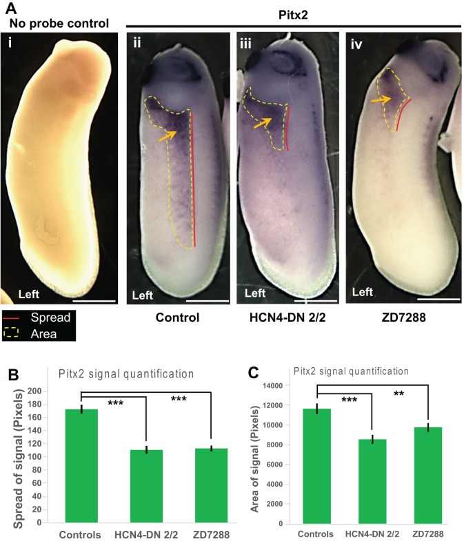
Pitx2 expression is affected by HCN4-DN and ZD7288. (A) Representative images of approximately stage 28 embryos assayed for Pitx2 expression by in situ hybridization. Left orientation of the embryo is indicated at the bottom of the image. Red line indicates the anterior-posterior spread of the Pitx2 expression and yellow dotted line indicates the area of the Pitx2 expression. (i) No probe (negative) untreated control, (ii) control embryos with Pitx2 signal – yellow arrow, (iii) embryos injected with HCN4-DN mRNA in both blastomeres at 2-cell stage with Pitx2 expression - yellow arrow, (iv) ZD7288-treated (100 µM stage1-10) embryo with Pitx2 expression - yellow arrow. Scale bar: 0.25 mm. (B) Quantification of anterior-posterior spread of Pitx2 expression (as indicated by red lines in A) in embryos showed a significant reduction in the spread of Pitx2 expression in HCN4-DN mRNA-injected and ZD7288-treated embryos. N=20; data was analyzed by one-way ANOVA; ***P<0.001. (C) Quantification of area of Pitx2 expression (as indicated by yellow dotted lines in A) in embryos showed a significant reduction in the area of Pitx2 expression in HCN4-DN mRNA-injected and ZD7288-treated embryos. N>25; data was analyzed by one-way ANOVA; ***P<0.001, **P<0.01.

