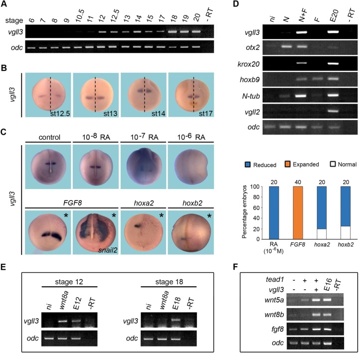
Fig. 1.
Temporal expression and spatial regulation of vgll3 in Xenopus embryo. (A) Vgll3 expression detected by RT-PCR starts between stages 11 and 12. (B) Vgll3 is detected by ISH in r2 during neural tube closure. Dashed lines indicate the midline of embryos. (C) Vgll3 expression decreases in stage 18 embryos treated with increasing concentrations of retinoic acid (RA). FGF8 mRNA-injected embryos show an anterior-lateral enlargement of vgll3 expression domain. Hoxa2 or hoxb2 mRNA-injected embryos show a strong reduction of vgll3 expression. All views are dorsal-anterior. Asterisks indicate the injected side. Quantification of vgll3 regulation results is shown in the right panel. Three independent experiments were performed. The number of embryos analyzed is indicated on the top of each bar. (D) Vgll3 is induced in animal caps treated with noggin+FGF2 (N+F). (E) Vgll3 expression is induced in early, but not late, animal cap cells overexpressing wnt8a. (F) Overexpression of vgll3 in combination with tead1 in animal cap cells stimulates the expression of wnt5a, wnt8b and fgf8. E, noninjected embryo (number indicates the stage); ni, animal cap from uninjected embryo; N-tub, N-tubulin; –RT, no cDNA. Ornithine decarboxylase (odc) gene expression was used as a control.

