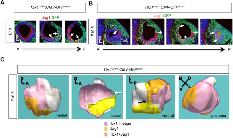
Fig. 2.
Tbx1 lineage is largely complementary to the NSD. (A,B) Immunofluorescence for Jag1 (red) and GFP (green) on transverse sections of a Tbx1Cre/+;CMV-GFP flox/+ embryo at E10 and E10.5, showing largely complementary expression between Jag1 and the Tbx1 cell lineage in more anterior sections of the OV, but some colocalization (white arrows) in more posterior ventral regions. (C) 3D reconstruction of serial sections of the E10.5, Tbx1Cre/+;CMV-GFP flox/+ embryo shown in Fig. 3C, showing the position of expression of the Tbx1 lineage and Jag1 as well as co-expression of both genes. The Tbx1 cell lineage is shown in pink, Jag1 protein expression is shown in yellow, and the overlap between the two is shown in orange. A dorsal-ventral (white arrow) and medial-lateral (black arrow) border between the Tbx1 lineage and Jag1 expression is shown.

